Pigmented Lesions & Melanoma Services
Our NYU Langone dermatologists are experts in the diagnosis of all skin cancers. We specialize in providing care for people who are at increased risk of developing melanoma, including people who have many pigmented lesions such as moles and atypical or dysplastic nevi, and people who have already had melanoma or who have a family history of the condition.
Our team has a long history of leadership in the diagnosis of melanoma at its early, surgically curable stage. Most notably, doctors at NYU Langone developed the “ABCDE” method for identifying melanoma, which is now widely recognized. We also pioneered the use of standardized total body photography to monitor people at increased risk for melanoma, and dermoscopy and other noninvasive methods to improve the accuracy of our diagnoses of skin lesions that are suspected of being cancer. Our dermatologists are leaders in educating dermatologists-in-training and dermatologists throughout the world in these methods.
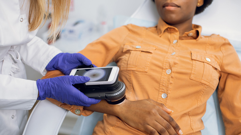
Our patients benefit from our use of the latest noninvasive diagnostic methods and tools. These include total body photography to monitor people who have an increased number of pigmented skin lesions, and dermoscopy, which uses a handheld magnifier that allows dermatologists to look deeply into the skin. We also use sequential digital dermoscopic monitoring, which allows us to detect skin cancer at the earliest stages while performing fewer biopsies of harmless lesions.
The latest addition to our noninvasive diagnostic toolkit is an imaging technology called reflectance confocal microscopy (RCM). RCM is a U.S. Food and Drug Administration–approved, noninvasive imaging tool that is designed to capture highly magnified images of the skin in real time, at a nearly microscopic level. It provides a more accurate diagnosis of melanoma than any other skin examination method. This leads to detection of very early stage melanomas, particularly on the face, and fewer biopsies of benign lesions.
RCM may also be used as a guide for biopsies of large, atypical lesions on the face that may be a symptom of lentigo maligna, a slow-growing form of melanoma found on skin that has received a lot of sun damage. This tool can also be used for presurgical imaging of skin cancers that have been already biopsied. This allows our surgeons to use smaller margins of normal skin when removing skin cancers, preventing unnecessarily large scars in areas where tissue preservation is of importance—for example, on the face, hands, feet, or the genitals—and reducing the risk of local recurrence.
When biopsy—the removal of a small skin sample—is necessary to analyze a suspicious lesion, our expert dermatopathologists provide quick and precise analysis, allowing you to receive an accurate diagnosis as soon as possible. Diagnosing and removing melanoma at its earliest stages offers you the best chance for a full recovery.
Our Pigmented Lesions and Melanoma Team
Our team of dermatologists has extensive experience in evaluating and diagnosing atypical nevi and melanoma. We collaborate actively with Perlmutter Cancer Center physicians as needed to provide the highest-quality care.