

Main content
Congenital heart defects occur when a baby’s heart does not form properly as it develops in the womb, causing a variety of conditions that affect heart function.
Babies with certain genetic conditions, such as Down syndrome, have a higher risk of developing a congenital heart defect, as do infants with a family history of congenital heart defects. Most often, though, the reason for the heart defect is unknown.
The symptoms of a congenital heart defect depend on the type of heart defect and the child’s age. Although a baby is born with a congenital heart defect, symptoms do not always appear immediately. In some children, symptoms that appear during infancy or even later may be the first sign of a problem.
Newborns with a congenital heart defect may have symptoms such as irritability or inconsolable crying, rapid breathing, excessive sweating, and difficulty feeding and gaining weight.
Symptoms in babies occur when the blood does not receive enough oxygen or the heart cannot pump efficiently. Symptoms often include: cyanosis, in which the skin appear bluish; fluid retention in the chest; a heart murmur, which the doctor can hear with a stethoscope; or an absent or rapid pulse. Decreased blood flow to the arms and legs may make a baby’s skin abnormally pale and cool.
In older children and adolescents, congenital heart defects may affect growth and development and produce weakness, fatigue, and shortness of breath during normal activities and exercise.
Specialists at the Pediatric Congenital Heart Program, part of Hassenfeld Children’s Hospital at NYU Langone, may diagnose congenital heart defects before birth, shortly after birth, or during childhood. They do so in collaboration with maternal–fetal medicine specialists and pediatricians.
A diagnosis of a congenital heart defect often begins when something unusual is noticed on a routine prenatal ultrasound, while a baby is in the womb. If a congenital heart defect is suspected on routine ultrasound, then the mother is referred to a pediatric cardiologist for a fetal echocardiogram.
Other prenatal tests may be recommended if a mother has a congenital heart condition or if the baby has other risk factors for congenital heart disease, including inherited conditions such as Down syndrome.
The earlier our team can identify a congenital heart condition, the better prepared our team can be to deliver treatment after the child is born.
After birth, heart specialists perform a physical exam and several other tests to confirm congenital heart disease in a newborn. These tests may also be used to make an initial diagnosis in infants and children who have symptoms of congenital heart disease but were not diagnosed prenatally.
During a physical exam, a doctor looks for signs that suggest your baby or child is not getting enough oxygen, such as bluish or unusually pale skin, lips, and nail beds; rapid breathing and flared nostrils; excessive sweating; an enlarged abdomen; and swelling of the fontanel, or “soft spot,” on the top of your baby’s head.
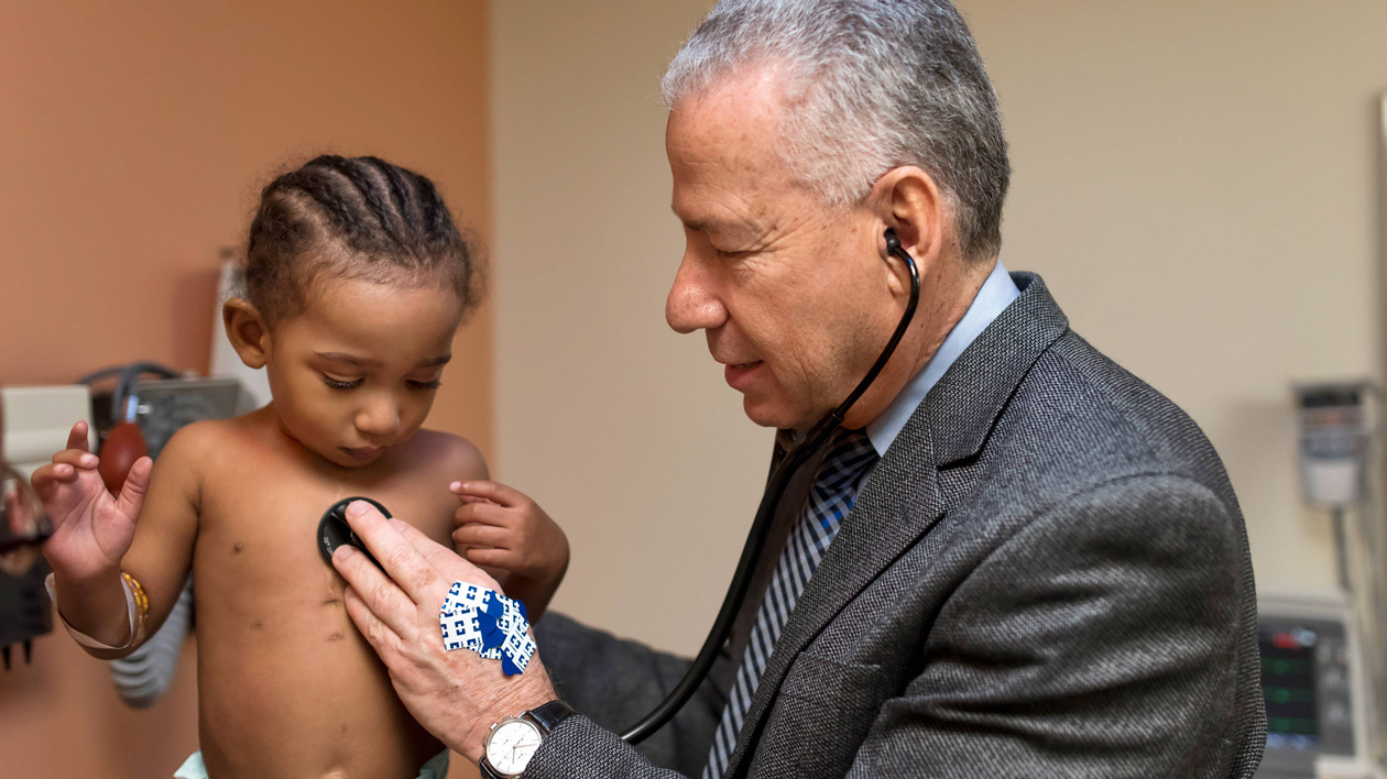
The doctor uses a stethoscope to listen for unusual heart sounds, such as clicks, irregular beats, or a murmur—a swishing sound that occurs when blood flows abnormally through a heart valve. The doctor also listens for breathing sounds, such as crackling, which may indicate a buildup of fluid in the lungs. Veins in the neck that are visible while your child is resting could be a sign that the heart isn’t pumping blood efficiently.
If your child is older, the doctor may ask you or your child about symptoms such as shortness of breath and fatigue during everyday activities or sports. The doctor also examines growth charts and other information from your pediatrician to see if your child is growing and developing as expected.
Specialists in our pediatric and fetal echocardiography laboratory are highly experienced in advanced cardiac imaging studies that identify pediatric heart defects.
An echocardiogram helps to evaluate the structure and function of the heart. In this test, an ultrasound wand called a transducer emits high-frequency sound waves that bounce off of organs and produce a precise computerized image of the heart, valves, and blood vessels.
A fetal echocardiogram, performed before birth, can be used to detect heart problems as early as the 18th week of pregnancy. During a fetal echocardiogram, a doctor or technician places the small ultrasound probe on the mother’s abdomen or inside the vagina. If the fetal echocardiogram shows that your baby has a heart condition, doctors from our fetal cardiology program coordinate the delivery and care of your newborn with your obstetrician and a team of neonatal heart specialists, including cardiologists, surgeons, and social workers.
In a baby or young child, a transesophageal echocardiogram produces images using an ultrasound probe inserted into your child’s throat and the esophagus, a part of which is located behind the heart. This method requires anesthesia.
This test allows doctors to view structures in the chest that a conventional echocardiogram doesn’t permit, and it gives detailed images of the heart. It’s also used during surgery and catheter procedures to help the surgeon see the area being repaired.
A three-dimensional echocardiogram generates a precise view of the heart and how it works. It provides views of the heart from all sides, which allows for accurate measurement of the size, shape, and position of heart defects, such as a hole or valve defect. Three-dimensional echocardiogram is often used in planning surgery.
An MRI scan uses a magnetic field and radio waves to generate detailed, cross-sectional images of the heart on a computer monitor, creating still and moving images of the heart. This scan can be used to evaluate the heart’s structure, its function, and blood flow to and from the heart. It can also measure the size of the heart’s chambers and major blood vessels. MRI can be used to detect signs of infection, tumors, or other heart conditions.
If your child is very young, an anesthesiologist may administer short-acting anesthesia to ensure that he or she remains still. During the test, your child lies on a narrow table that slides into the hollow, tube-shaped scanner. The scanner makes loud banging noises, which can be blocked out with special headphones.
MRI may be performed with or without contrast dye, which is injected into a vein before the test and allows the doctor to see the images in greater detail. The test is usually completed in about an hour.
Information from CT and MRI scans may be used to create a three-dimensional heart model to plan surgery or a catheter procedure. A three-dimensional heart model is an exact replica of your child’s heart, created using the imaging tests and a three-dimensional printer. The model, made of a special type of plastic, allows the doctor to view the location and size of a heart defect, which can help to determine the best approach to treatment.
A CT scanner uses beams of radiation and moves in a circle around the body. Information from these X-rays is transmitted to a computer that forms a detailed image.
As with an MRI scan, your child may be given a mild sedative to help him or her to stay still during this test. Your child lies on a narrow table that slides into a doughnut-shaped hole inside the scanner. CT scans may be performed with or without contrast dye and take only a few minutes.
Cardiac catheterization is a minimally invasive procedure that is often used to diagnose congenital heart defects after birth. Learn more about cardiac catheterization.
An electrocardiogram, sometimes called an EKG, records electrical activity in the heart. An electrocardiogram can help to identify an abnormal heart rhythm caused by a congenital heart defect. This test is also performed during and after catheter procedures and open heart surgery.
Before an electrocardiogram, a technician places 12 stickers that contain small electrodes on your child’s chest, arms, and legs. The electrodes are painless, so no sedation is needed. The electrodes transmit electrical impulses to a machine that traces electrical activity in the heart within minutes.
Depending on the way the tracing appears, the electrocardiogram can help the doctor to determine if your child’s symptoms are caused by damage to the heart’s electrical system, abnormal blood flow to the coronary arteries, an electrolyte imbalance, or inflammation in the heart.
Holter and event monitors are portable devices used to evaluate symptoms such as dizziness, fainting, low blood pressure, chest pain, and heart palpitations. Normally, heart rhythm varies throughout the day, depending on a person’s activities. Unlike an electrocardiogram, which takes a snapshot of the electrical activity in the heart at one brief point in time, this monitoring shows changes in heart rhythm over a longer period of time.
The Holter monitor records your child’s heart rhythm over a 24-hour period, during everyday activities, including sleep. The device contains a small, rectangular monitor that can be worn around the waist and is connected to five small electrodes attached to your child’s chest.
An event monitor measures electrical activity for one to several days. Unlike the Holter monitor, which records electrical activity continuously, the event monitor records electrical activity only when your child feels symptoms. A technician explains how and when to push a button that instructs the monitor to record electrical activity and how to send the data to the doctor’s office. You or your child may also write down what your child was doing when symptoms began, which can help to determine if there is a link between activities and symptoms.
A stress test may be used to see how the heart of an older child or adolescent responds to exercise, such as riding a stationary bicycle or walking on a treadmill. The placement of electrodes used in a conventional electrocardiogram is modified to allow for movement of your child’s arms and legs.
Before starting the exercise, the technician performs an initial electrocardiogram and measures your child’s blood pressure with a blood pressure cuff. Blood pressure and electrical activity are monitored as your child begins to exercise and as the exercise level gradually increases during the 30-minute test. A reading is taken about 15 minutes after your child stops the exercise.