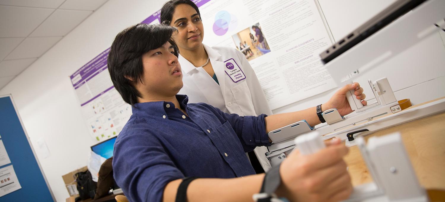
Preeti Raghavan, MD, working with a research assistant on the m2 BAT device.
Photo: NYU Langone Staff
There is no better example of the way research informs care at Rusk Rehabilitation than the m2Bimanual Arm Trainer (BAT), the invention of Preeti Raghavan, MD, director of NYU Langone’s Motor Recovery Research Laboratory.
Dr. Raghavan led development efforts for a range of patented motor rehabilitation devices now being used at Rusk and elsewhere.
The m2 BAT is based on the principle of “mirrored motion,” which stimulates the two sides of the brain to work together to restore movement in the affected arm after a stroke. This year, the m2 BAT was cleared by the FDA as a Class 1 device and is now being used by Rusk therapists in stroke rehabilitation. What makes the m2 BAT special is a technological leap that uses a video game interface to provide a virtual, programmable training experience that can be individualized to the patients’ stage of recovery. Furthermore, real-time feedback of the movement motivates patients to push beyond their current abilities, while the device guides them to use the right movements. Dr. Raghavan is now working with colleagues to explore the use of the m2 BAT as a home-based rehabilitation tool, with treatment monitored remotely by the therapist—exciting progress toward the goal of home-based personalized telerehabilitation.
Dr. Raghavan and her team are also currently working under a prestigious 5-year R01 NIH research grant to develop innovative approaches to restore dexterous hand function. A hallmark of dexterity is the ability to quickly learn to use one’s senses, such as touch, vision, and kinesthetic sense from muscles, to produce finely tuned movements and forces that are appropriate for each task. Recent results published in the Journal of Neurophysiology and presented at the Society for Neuroscience meeting show how the fingertip forces and muscle activity patterns are finely tuned to specific types of sensory information. Since individuals with stroke may have varying degrees of sensory impairment, these results will help understand why dexterity is impaired in a given patient and what can be done to restore it. Dr. Raghavan and her team are focusing on developing an algorithm that will inform the appropriate rehabilitation strategy for patients with different sensory impairments.
Investigating a Multisensory Approach to Rehabilitation
With his team, John-Ross (J.R.) Rizzo, MD, director of the Visuomotor Integration Laboratory (VMIL) and assistant professor of rehabilitation medicine and neurology, focuses on taking a multisensory approach to stroke care. Dr. Rizzo published research on what is known as the dual-task phenomenon, where patients carry out motor and cognitive tasks simultaneously, engaging multiple sensorimotor systems. One study, which appeared in the April 2015 issue of the Archives of Physical Medicine and Rehabilitation, involved subjects walking while listening to emotionally salient sounds that typically demand increased attention. Dr. Rizzo and colleagues used a cognitive assessment tool known as the Blessed Dementia Scale (BDS) to grade the mental ability of subjects and found that for subjects at the upper limit of normal on the BDS (those close to cognitive impairment score cutoffs) had a slower gait speed during the dual task, as compared to healthy age-matched controls. Results from this pilot study suggest that such a dual-task assessment might be useful as a tool for screening of cognitive impairment—one that might lead to earlier and more effective interventions.
Also, Dr. Rizzo’s research into how stroke affects control of eye movements may revolutionize the approach to rehabilitation after stroke. Using sophisticated camera technology, researchers record the eye movements of stroke patients in fine detail. Subjects are asked to follow targets that move around very quickly on a computer screen. The test measures saccadic eye movements, a physiologic event in which an individual quickly moves his gaze from one point of interest to another. Dr. Rizzo and his team expected to find that despite normal visual evaluations, stroke patients would have impaired saccadic eye movements. Specifically, the team hypothesized that the eye movements would be slowed. However, they found the opposite. Subjects exhibited faster initiation times of saccadic eye movements. Dr. Rizzo and the team have hypothesized that these findings suggest a disinhibition phenomenon, akin to other phenomena after stroke such as hyperreflexia and spasticity, that suggest impairment in the cortical braking system. The extent of impairment in eye movement control may reflect the state of the neural systems and inform rehabilitation strategies.

