The Two Diseases Cause Damage in Some of the Same Ways
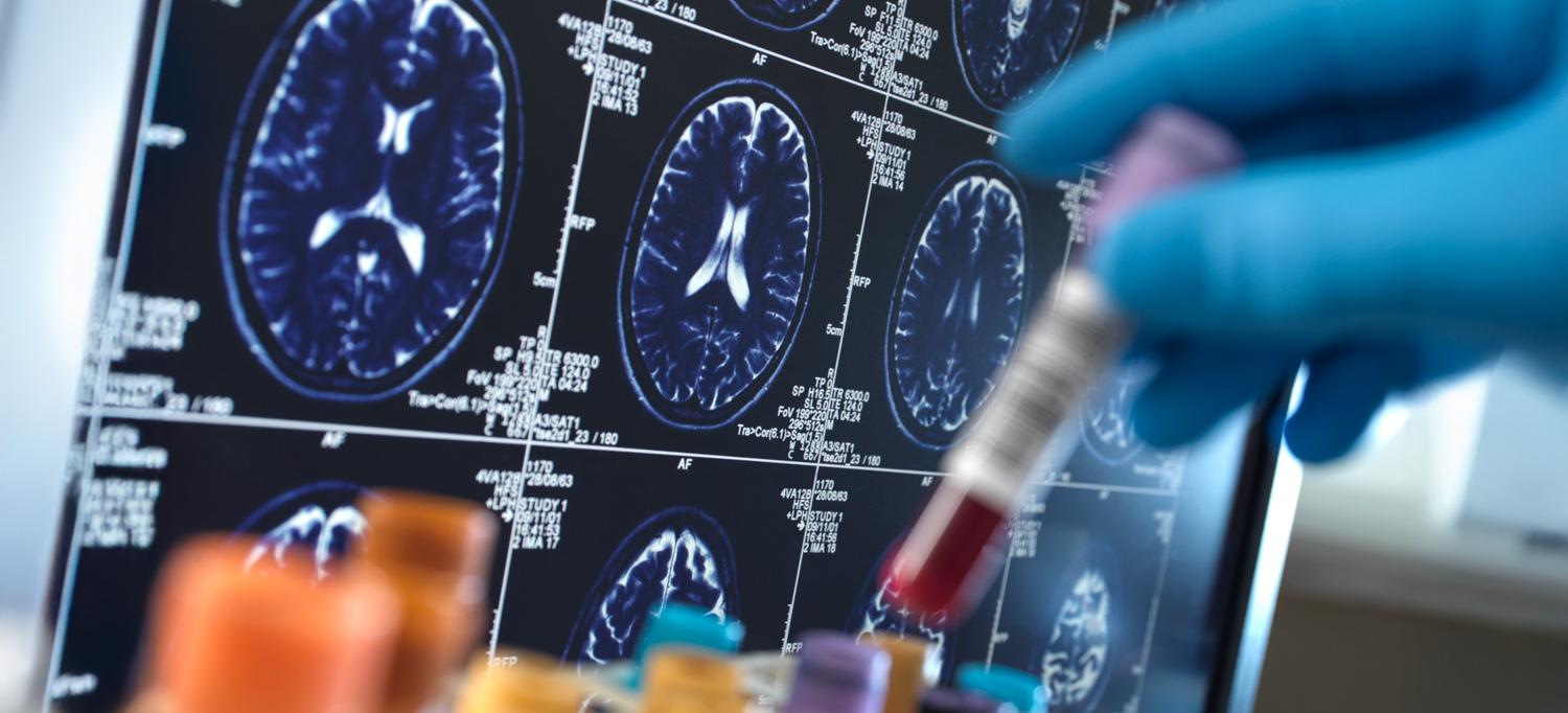
Study participants with symptoms of long COVID had a brain structure that was 10 percent larger than those who had fully recovered.
Credit: Getty / Andrew Brookes
The increased size of, and reduced blood flow to, a key brain structure in patients with long COVID tracks with known blood markers of Alzheimer’s disease and greater levels of dementia, a new study found.
Led by researchers at NYU Langone Health, the study concerns the choroid plexus (CP), a network of blood vessels lined by cells that produce cerebrospinal fluid, which cushions the brain and forms a protective barrier between the fluid and the bloodstream. The CP is known to regulate immune system responses (inflammation) and waste clearance in the brain, with past studies showing that the COVID virus can damage the cells that line CP blood vessels.
Published online recently in Alzheimer’s & Dementia, the journal of the Alzheimer’s Association, the new work found that patients reporting long COVID had a 10 percent larger CP than those who had fully recovered from an initial infection with SARS-CoV-2, the virus that causes COVID-19 disease. Further, CP size increases tracked with blood levels of proteins known to increase as Alzheimer’s disease worsens, like pTau217, and with others that rise in response to brain injury, like Glial fibrillary acidic protein (GFAP).
The research team also found that those patients with larger CPs performed an average of 2 percent worse on a standard 30-point screening test, the mini–mental state examination (MMSE), which measures changes in memory and attention.
“Our work suggests that long-term immune reactions caused in some cases after an initial COVID infection may come with swelling that damages a critical brain barrier in the choroid plexus,” said senior study author Yulin Ge, MD, a professor in the Department of Radiology at NYU Grossman School of Medicine. “Physical, molecular, and clinical evidence suggests that a larger CP may be an early warning sign of future Alzheimer’s-like cognitive decline.”
Long COVID, sometimes called post-acute sequelae of COVID-19 (PASC), is a condition in which symptoms of infection with the pandemic virus last for months or even years after the initial infection with SARS-CoV-2. About 780 million people worldwide have been infected with the virus so far, with some of those infected experiencing long-term fatigue, brain fog, dizziness, loss of smell or taste, depression, and many other symptoms.
Linking Changes
For the study, the research team recruited 179 participants: 86 patients with neurological symptoms of long COVID, 67 people who had fully recovered from COVID-19 without lasting symptoms, and 26 healthy individuals who had never had COVID-19.
Each participant underwent advanced MRI scans of their brains, which enabled the team to find patterns amid the brain scans, blood tests, and cognitive exams of the three patient groups.
The tests revealed that structural changes seen in the CP blood vessels of the long COVID patients both expanded CP volume and reduced flow through its blood vessels. Although the mechanisms behind this are not yet confirmed, the team’s theory is that the changes reflect inflammation-driven CP “vascular remodeling,” in which layers of cells lining blood vessels thicken in response to long-term activation by immune cells and signals. Such inflammation comes with surrounding stromal fibrosis, the buildup of scar-like tissue that further hinders blood flow.
Impaired blood perfusion in the CP may hinder production of cerebrospinal fluid, lead to waste buildup, and compromise the integrity of the blood–fluid barrier, the study authors said.
“Our next step is to follow these patients over time to see if the brain changes we identified can predict who will develop long-term cognitive issues,” said senior study author Thomas M. Wisniewski, MD, the Gerald J. and Dorothy R. Friedman Professor of the New York University Alzheimer’s Disease Research Center, which is part of the Department of Neurology at NYU Grossman School of Medicine. “A larger, long-term study will be needed to clarify whether these CP alterations are a cause or a consequence of the neurological symptoms, which promises to better focus treatment design efforts,” added Dr. Wisniewski, who is also director of the Center for Cognitive Neurology at NYU Langone.
Along with Dr. Ge, study authors from the Department of Radiology were Huize Pang, who was first author; Li Jiang; Chenyang Li; and Zhe Sun. In addition to Dr. Wisniewski, authors from the Department of Neurology were Jennifer A. Frontera, MD; Allal Boutajangout, PhD; Ludovic Debure; Mobeena Ghuman; Alok Vedvyas; and Arjun V. Masurkar, MD, PhD.
This research was supported by National Institute on Aging grants R01AG077422, U24 NS135568, and P30AG066512.
About NYU Langone Health
NYU Langone Health is a fully integrated health system that consistently achieves the best patient outcomes through a rigorous focus on quality that has resulted in some of the lowest mortality rates in the nation. Vizient, Inc., has ranked NYU Langone No. 1 out of 118 comprehensive academic medical centers across the nation for four years in a row, and U.S. News & World Report recently ranked four of its clinical specialties No. 1 in the nation. NYU Langone offers a comprehensive range of medical services with one high standard of care across seven inpatient locations, its Perlmutter Cancer Center, and more than 320 outpatient locations in the New York area and Florida. The system also includes two tuition-free medical schools, in Manhattan and on Long Island, and a vast research enterprise.
Media Inquiries
Greg Williams
Phone: 212-404-3500
Gregory.Williams@NYULangone.org