Photo: PhotoAlto/Michele Constantini/Getty
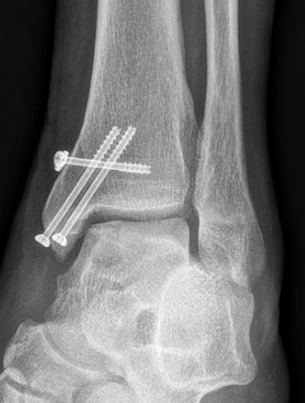
A highly experienced foot and ankle surgeon, an unusual surgical approach, and multidisciplinary expertise at NYU Langone combined to help a former athlete and paratrooper overcome a severe ankle injury. The 55-year-old patient presented with progressively worsening and debilitating pain in his ankle. An initial MRI showed a large osteochondral lesion (OCL) in his medial talar dome with uncontained, multiloculated cysts.
Estimates suggest that up to half of all acute ankle sprains and fractures include some form of osteochondral injury. The patient, told by other doctors that he would need an ankle fusion or replacement, came to NYU Langone because of its expertise in treating such OCLs.
An OCL of the talus can be surgically approached in two ways: by either repairing or replacing the damaged cartilage. “Lesion size is important to determining the procedure,” says John G. Kennedy, MD, professor of orthopedic surgery and chief of the Division of Foot and Ankle Surgery at NYU Langone. If the lesion is less than 1 centimeter, for instance, surgeons often recommend a reparative technique that includes bone marrow stimulation. “On the other hand, if the lesion size exceeds 1 centimeter, we often apply a replacement technique that includes autologous osteochondral transplantation, or AOT,” Dr. Kennedy says.
A cystic lesion and uncontained lesion are both prognostic indicators for poor outcomes of the microfracture-based reparative procedure, according to guidelines made in 2017 at a consensus meeting in Pittsburgh held by the International Society on Cartilage Repair of the Ankle (ISCRA), which Dr. Kennedy cofounded.
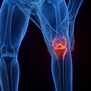
Since the patient’s lesion was larger than 1 centimeter, he instead required an AOT replacement procedure, which involved using two cylindrical osteochondral grafts from his knee. “Typically we use one graft,” Dr. Kennedy says. “But in this case, since the lesion size was large, we performed double grafts.” This double-plug AOT technique is not only rarely performed but also highly dependent on multidisciplinary collaboration with radiology and other departments.
Dr. Kennedy was greatly aided in his surgery by MRI T2 mapping of the patient’s ankle cartilage. Yearly, high-resolution images using the same MRI technique can help confirm both the quality and quantity of the cartilage transplant and allow the surgical team to follow patients carefully. Typically, impending cartilage issues can be addressed nonsurgically with biologic injections of growth factors that help preserve and protect new cartilage formation.
Dr. Kennedy has performed the AOT procedure, one of his specialties, more than any other orthopedic surgeon in the country. For this surgery, he also used concentrated bone marrow aspirate (CBMA), which he harvested from the patient’s iliac crest via a centrifuge system. “CBMA has stem cells and lots of growth factors and potent anti-inflammatory protein that can help with the incorporation of cartilage and bone, and reduce inflammation,” Dr. Kennedy says. In addition, CBMA can reduce the cyst occurrence rate after the AOT procedure, making it a key part of greatly improving clinical outcomes.
The challenging AOT surgical procedure requires perfect alignment; the cutting-edge combination therapy, though, can allow more than 90 percent of athletes to return to their previous level of competitive sports. The precision technique, high-end imaging, and experienced surgeon and surgical team yielded an outstanding outcome for the former paratrooper. Three months after the surgery, with physical therapists guiding his recovery, he was able to return to his activities.


