Photo: SCIENCE PHOTO LIBRARY/Getty
A common side effect of radiation therapy for breast cancer and other cancers is a reddening of the skin called radiation dermatitis. Doctors typically grade the severity of this skin condition on a scale of 1 to 4, with 4 being the most severe. However, the grading system is subjective—physicians have different thresholds for what they observe to be a grade 1 to 4 dermatitis—and the physician-graded scale does not take into account variability in skin of color.
A recently launched study at NYU Langone Health’s Perlmutter Cancer Center aims to measure radiation dermatitis in women receiving breast radiation to better understand radiation dermatitis in women with skin of color.
The study, a single-arm, institutional, nontherapeutic trial, was conceived and designed by Juhi Purswani, MD, a postgraduate year 4 resident in the Department of Radiation Oncology at NYU Grossman School of Medicine, under the mentorship and guidance of Naamit K. Gerber, MD, a radiation oncologist at Perlmutter Cancer Center and director of the Radiation Oncology Residency at NYU Grossman School of Medicine.
“Some patients who have darker pigmented skin become tan or acutely hyperpigmented—not red—when they have been exposed to the ionizing radiation used to treat cancer,” Dr. Purswani says. “That color change is not captured in the scale. We think radiation dermatitis is underdiagnosed in those patients because we are not considering acute tanning of the skin to be inflammation related to radiation treatment.”
Dr. Gerber, principal investigator of the study, plans to enroll 80 women who are candidates for partial breast, whole breast, or chest wall radiation therapy following breast-conserving surgery or a mastectomy. The study will follow participants receiving three radiation therapy regimens: ultra-hypofractionation, which is completed after one week of radiation; hyperfractionation, which is given over three to four weeks of radiation; and conventional radiation therapy, which is given over five to six weeks.
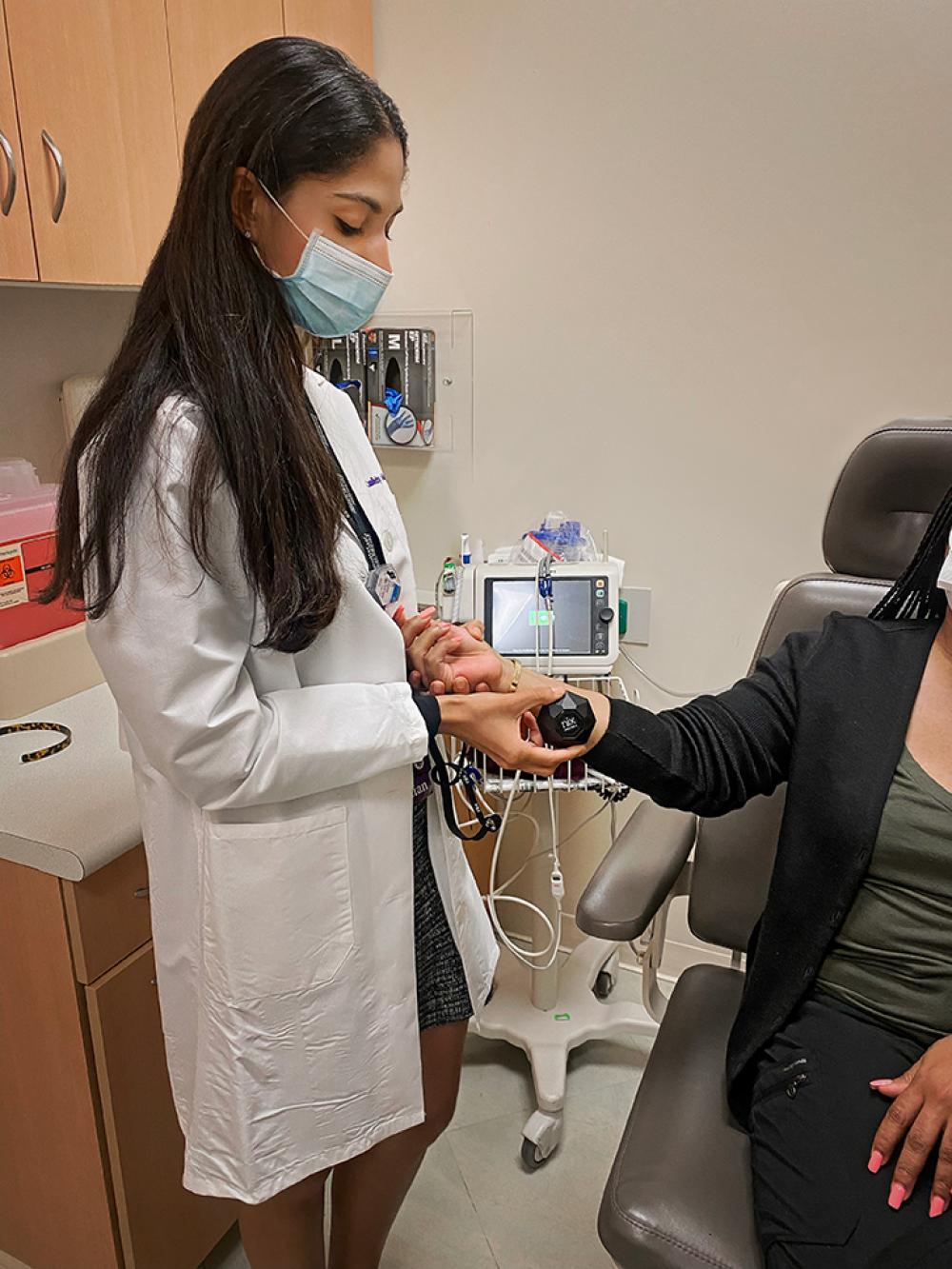
Using a portable spectrophotometer, a device commonly used in the cosmetic and paint industries to match colors, the research team will measure participants’ baseline skin color before receiving radiation therapy. The spectrophotometer shines stable, broad-spectrum white light over the skin and collects and processes spectral data from the light reflected back from the skin.
The study is focusing on three key numeric values that produced by the device: L, which is a measure of pigmentation change on a black–white scale; A, which is a measure of superficial reddening of the skin called erythema on a red–green scale; and B, which is a measure of the blue–yellow color scale. The L value measures the degree of hyperpigmentation, and the A value provides a measure of erythema or skin reddening.
Skin color will be measured once a week during radiation therapy, and then at two weeks, one month, and one year after the completion of radiation therapy.
“We are using the spectrophotometer to track the changes that are occurring at predetermined locations on the breast or chest wall so that we can look at these objective numeric parameters that are related to the skin that capture both reddening of the skin and pigmentation change.” Dr. Purswani says. “By capturing these changes objectively and longitudinally throughout treatment, we can look for trends that are unique to patients based on their baseline skin tone and identify opportunities for earlier intervention and individually tailored care for the patient.”
The study is funded by a Radiation Oncology Resident Seed Grant from the American Society for Radiation Oncology (ASTRO) and the Breast Cancer Research Foundation.
“This is an exciting study for breast radiation oncology because we do not have adequate tools to characterize and quantify radiation dermatitis in patients with skin of color,” says Dr. Gerber, also an associate professor in the Department of Radiation Oncology at NYU Grossman School of Medicine. “Dr. Purswani’s project is exploring a little-known area of radiation oncology, and she serves as a model for the ways in which resident physicians can conduct groundbreaking research.”


