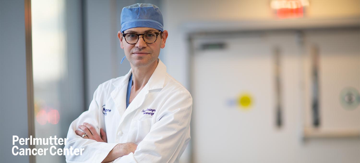
Dr. Daniel A. Orringer has developed a self-contained AI platform, called DeepGlioma, that can be wheeled into an operating room to speed up analysis of tumor specimens.
Photo: NYU Langone Staff
For more than a century, pathologists have used microscopes to study the size, shape, and arrangement of cells in thin sections of human tissue to help diagnose disease and guide clinical decision-making. Yet, looking at the size and shape of cells—a field called histology—only tells part of the story. And because pathologists vary in their interpretation of data, it’s hard for humans to consistently make accurate diagnoses based on analysis of images alone. This can lead to confusion about disease prognosis and the best treatment approach. Increasingly, pathologists rely on molecular testing to assist in diagnosis when the histology is unclear. However, molecular testing is costly, time-intensive, and not available in all settings where patients with cancer are treated.
Daniel A. Orringer, MD, a neurosurgeon at NYU Langone Health’s Perlmutter Cancer Center, is developing artificial intelligence (AI)–based tools to facilitate histologic and molecular diagnosis and personalize treatment for people with brain tumors.
“We have been able to teach an AI algorithm to detect features within microscopic images of the tissue, and, in a systematic way, to infer the molecular diagnosis of a brain tumor just from the image,” said Dr. Orringer, who is an associate professor in the Departments of Neurosurgery and Pathology at NYU Grossman School of Medicine and a member of the cancer center’s Brain and Spine Tumor Center. “We have trained an algorithm to forecast molecular alterations within tumor cells, without the need for mutational sequencing or chromosomal analysis, to quickly tailor treatment.”
For more than a decade, physicians caring for patients with brain tumors have used molecular classification to standardize diagnosis. One of the most common types of tumors that form in the brain are gliomas, almost all of which are defined by a handful of genetic alterations.
Pathologists, however, can’t determine these genetic alterations by looking at traditional slides. That’s where AI comes into play.
In a study published March 23 in Nature Medicine, Dr. Orringer and his colleagues showed that an AI-based screening system called DeepGlioma could analyze tumor specimens taken during an operation and detect genetic mutations in diffuse gliomas in less than 90 seconds.
“DeepGlioma is a game changer for patients, primarily because the molecular testing that is normally required to be able to arrive at a diagnosis, even in the best resourced centers, can take weeks to come back,” Dr. Orringer said. “Instead of waiting weeks to know what kind of glioma a patient has and how to treat it, we get that information in a matter of minutes. This expedites care and allows the surgeon in the operating room to tailor the surgical approach.”
The type of tumor a patient has dictates the surgical approach, whether it should be more conservative or more aggressive. Knowing the molecular classification of the tumor also allows the medical and radiation oncologists to choose the most appropriate type of therapy.
There are two major types of gliomas: those with mutations of the IDH gene and those without IDH mutations. The presence of IDH mutations generally correlates with a better prognosis compared to gliomas without IDH mutations, but there is also variability among IDH-mutant gliomas.
IDH mutant gliomas are divided into two major categories: astrocytoma (IDH mutant, 1p19q intact) and oligodendroglioma (IDH mutant, 1p19q co-deleted). The prognosis and response to surgery, chemotherapy, and radiation vary dramatically amongst the two major types of IDH-mutant gliomas.
Oligodendrogliomas are highly sensitive to standard chemotherapy and radiation and carry a better prognosis regardless of how much tumor is removed at the time of surgery.
Astrocytomas are less responsive to chemotherapy and radiation. In addition, the percentage of tumor removed during surgery has a dramatic impact on patient outcome.
“We have been able to teach the algorithm to differentiate between the two types of IDH-mutant gliomas,” said Dr. Orringer. “We now know that surgical strategy should be altered based on genetic subtype in IDH-mutant gliomas. To date, we have had no way of knowing the genetics of tumors we’re operating on. Without real-time genetic testing, it’s not possible to know how aggressive we should be surgically and whether a patient is likely to respond to conventional chemotherapy and radiation.”
DeepGlioma unlocks the genetic makeup of gliomas in minutes in the operating room. Doctors can use this information to tailor a patient’s treatment to the molecular subtype of tumor that they have—a feat that would be impossible to do without the help of rapid AI-powered optical histology.
“Many patients who have brain tumors are treated in centers that are under-resourced with respect to molecular diagnostics. The system we developed doesn’t require the facilities within a pathology lab,” Dr. Orringer said. “Our platform for DeepGlioma is self-contained and can be rolled into any operating room; all that is needed is a plug. Not tissue processing or staining is required. Surgeons can go from fresh tissue to molecular forecasting without the need for any dyes or labels or processing of the tissue.”
Dr. Orringer envisions DeepGlioma as part of a screening system for molecular diagnosis. He and his team have been validating it at Perlmutter Cancer Center’s Manhattan sites to tailor surgical care. The system’s portability means that the technology will eventually be available across the cancer center’s network sites in Brooklyn and on Long Island. Dr. Orringer said that his team is also developing the system behind DeepGlioma in cancers other than glioma.
“The use of optical histology and AI to diagnose cancer will have implications for the way that many types of cancers are treated, not just those in the brain, but also those in the lung, breast, and prostate,” Dr. Orringer said. “People with cancer who receive treatment in a setting that has access to the most rapid and readily available molecular diagnostics have the potential to get a higher standard of care.”
Learn more from NBC News and Fox 5 New York.

