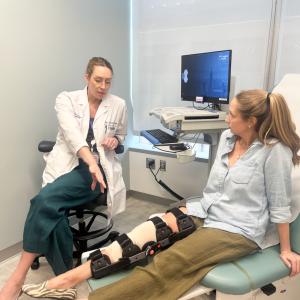
Dr. Aaron J. Buckland and his team developed a dislocation risk prediction model to screen patients undergoing hip replacement.
Photo: NYU Langone Staff
Researchers at NYU Langone Health have identified several new risk factors for implant dislocation following total hip replacement surgery, informing the development of a new preoperative screening tool and treatment algorithm to better identify patients with high risk based on these factors.
According to Aaron J. Buckland, MD, assistant professor of orthopedic surgery, the research is rooted in a growing understanding of the role of the spine in hip implant stability.
“In the last few years, we have learned that spinal pathologies markedly change the way the pelvis moves during posture changes,” says Dr. Buckland. “For example, people with spinal pathologies display much less posterior pelvic tilt when they go from standing to sitting.”
This effect is notable in patients with spinal degeneration or spinal deformity and in patients who have undergone previous fusion surgeries. “By reducing the change in posterior pelvic tilt in sitting, these conditions lead to a less protective anteversion acetabular component,” explains Dr. Buckland. “When this happens, it increases the risk of impingement and subsequent implant dislocation.”
Calculating the Risk of Hip Implant Dislocation
To explore the impact of spinopelvic mechanics on hip implant stability, Dr. Buckland and Jonathan Vigdorchik, MD, led a study analyzing 1,082 patients who had total hip replacement surgery in 2014 or 2015. The team identified an overall implant dislocation rate for this group of 1.8 percent; among the 320 patients demonstrating evidence of lumbar spinal degeneration or deformity, the dislocation rate was 3.1 percent.
Based on this data, the researchers developed a dislocation risk prediction model, used prospectively to screen patients undergoing hip replacement in 2016. Out of a total of 1,009 patients, 192 individuals were identified as high risk and were treated under a high-risk algorithm.
“In the group of patients who were put into our high-risk pathway, there was only one dislocation, which suggests a rate of 0.5 percent,” Dr. Buckland notes. “Compared to the previous group of patients with spinal pathologies treated with traditional strategies, these results represent a sixfold decrease in the dislocation rate.”
Aligning Risk with New Clinical Pathway for People Who Require Hip Arthroplasty
All patients who undergo hip arthroplasty at NYU Langone are now screened with the new risk assessment tool, which then assigns patients to risk categories based on spinal pathology and other recognized risk factors.
“Patients with the highest risk are those with lumbar severe spinal degeneration, lumbar flat back deformity, and those who have multiple lumbar fusion levels,” says Dr. Buckland.
The screening also takes into account known dislocation risk factors, including age greater than 75 years, female sex, diagnoses such as femoral neck fracture or avascular necrosis, and neurological diseases like Parkinson’s or dementia. “These are all intermediate risk factors, but a combination of them can make you a patient with high risk,” says Dr. Buckland. “This group also includes patients who engage in activities that put their hips into more extreme ranges of motion, like yoga instructors or surfers.”
Patients categorized as high risk are automatically assigned to the special treatment algorithm, a pathway that begins with a nonstandard imaging plan that more accurately assesses spinopelvic function. While traditional preoperative imaging for hip patients comprises a single anteroposterior radiograph in the supine position, patients with high risk receive sitting and standing X-rays, in both the anteroposterior and lateral views, under the new algorithm. “These additional views provide critical information on functional position, such as how much the pelvis tilts posterior when the patient sits,” says Dr. Buckland.
Radiographic assessment then informs hardware choice. “If the sitting and standing radiographs show very little leeway in both directions, we use a dual mobility implant,” Dr. Buckland continues. “The dual mobility bearing allows increased range of motion and gives the hip replacement more inherent stability.”
The high-risk algorithm also includes advanced intraoperative technologies. “The imaging studies help us plan the optimal position of the cup within the acetabulum, but then we still need to make sure we can achieve that placement during surgery,” says Dr. Buckland. “Tools like intraoperative navigation, laser guidance, and robotics help us place the implant precisely where we want it.”

