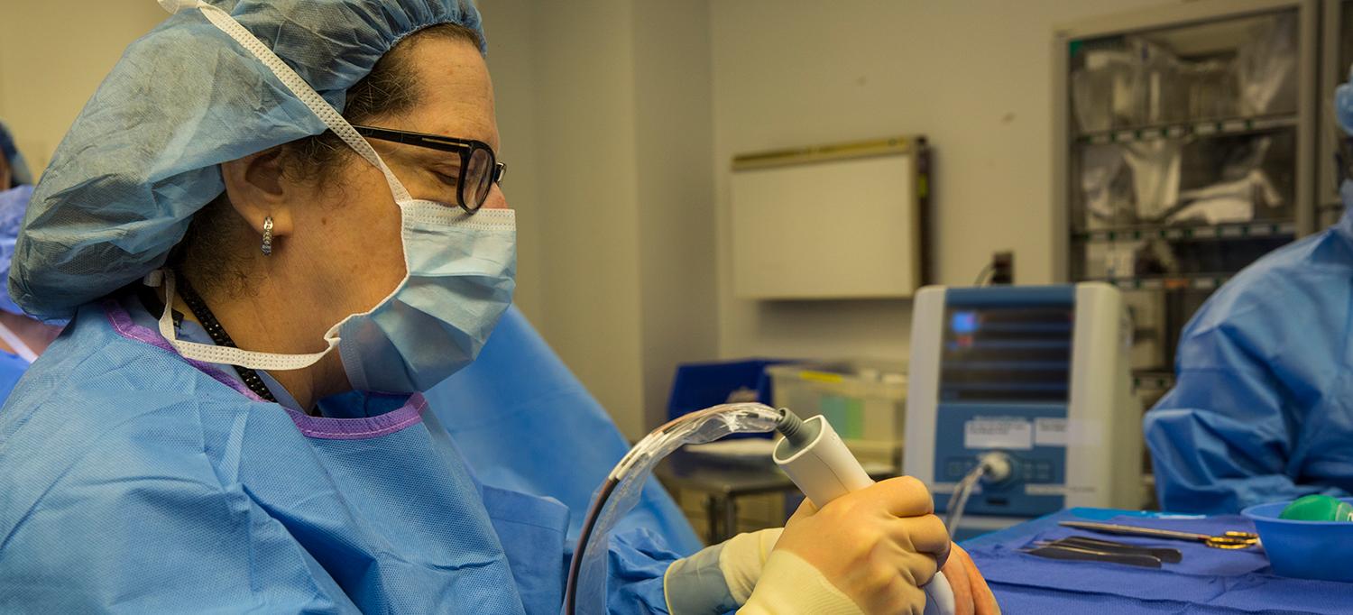
Dr. Freya Schnabel, director of breast surgery at NYU Langone.
Photo: Adam Watt
Whenever a breast cancer surgeon performs a lumpectomy, there’s always a lingering concern: Has enough surrounding tissue been removed to ensure that no malignant cells remain? Though surgeons have some tools to guide them, the only way to know for certain is for a pathologist to examine the margins, or edges, of the excised tumor following surgery. But thanks to a new device called MarginProbe®, which detects electromagnetic differences between breast cancer cells and normal breast tissue, surgeons can now get a more accurate assessment while they operate, sparing many women with early-stage breast cancer from additional surgeries.
NYU Langone was the first hospital in the tri-state area to use MarginProbe®, and our physician-researchers were part of a pivotal study of over 600 women that led to its approval by the FDA. The results showed that MarginProbe® was up to three times as effective in finding additional cancer on the margins of tumorous tissue as traditional methods, such as inspecting and imaging the tissue. It’s an advance that could benefit the more than 170,000 women who undergo a lumpectomy each year. “In about 20 percent of cases, surgeons find that they have to reoperate to remove additional tissue,” notes lead author Freya Schnabel, MD, director of breast surgery at NYU Langone’s Laura and Isaac Perlmutter Cancer Center. “We felt we could and should do better for our patients.”
Re-excision surgery is frustrating and stressful for the patient, Dr. Schnabel explains, and it may delay necessary follow-up treatments like radiation and chemotherapy. An additional operation can also result in negative cosmetic effects. “MarginProbe® is a major advance,” says Dr. Schnabel, because doctors and patients alike can feel more confident that only one surgery will be necessary.

