Anthony K. Frempong-Boadu, MD
PHOTO: Karsten Moran
For a 29-year-old patient presenting with a rare and risky intramedullary cavernoma spinal cord tumor, the use of advanced diffusion tensor imaging (DTI), pioneered at NYU Langone for spinal surgery, proved essential to a successful resection—and restored quality of life.
When Anthony K. Frempong Boadu, MD, clinical associate professor of neurosurgery and director of the Division of Spinal Surgery, evaluated the patient’s symptoms and progressing condition, he quickly saw the dilemma: Resection of such a complex tumor required the use of a leading-edge, highly precise imaging technique but one with only a limited track record—but postponing the surgery was not an option. The debilitating effects of the intradural intramedullary cavernoma had become increasingly apparent in the year since it was first diagnosed and they would only worsen as the tumor grew. “It was clear that this tumor needed to be managed surgically,” says Dr. Frempong-Boadu.
However, such a resection carried formidable risks. Intramedullary tumors, which account for just 2 to 4 percent of all intrinsic central nervous system tumors, often permeate the surrounding tissues after arising from the cells of the spinal cord. Resection of this tumor would require myelotomy—dissection of the spinal cord at the midline—to achieve access, a surgery with a high risk of permanent morbidity, damage to paraspinal structures, cerebrospinal fluid leak, central nervous system infection, and significant neurological injury, including paraplegia. The tumor was densely adhered to surrounding nerve tissue, increasing the likelihood of surgical complications. Other leading tertiary centers had been reluctant to perform the operation.
Advanced Neural Images Provide a Road Map for Resection
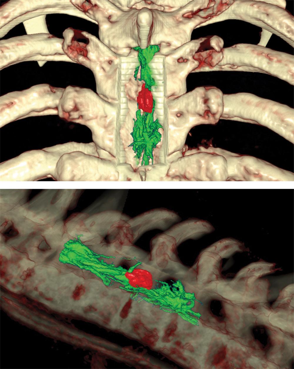
With Dr. Frempong-Boadu’s successful DTI application two years earlier, NYU Langone had become the nation’s only center to use real-time intraoperative functional data during spinal surgery. In this case, use of this approach—which employs multiple high-information MRI scans of the water diffusion rates across nerve membranes to map the exact location of a patient’s nerve tracks—was critical to keeping the patient neurologically intact as the tumor was resected.
“With DTI, combined with Surgical Theater’s three-dimensional virtual imagery of the surgical site, we could visualize exactly where the motor tracks of the patient’s spinal cord and all other relevant structures were at all times, so we could go in and confidently remove the tumor,” says Dr. Frempong-Boadu.
A Multidisciplinary Team Effort Augments Advanced Imaging
With the full array of spinal imaging techniques at work in the operating room, the surgical team began with a decompressive osteoplastic laminectomy of the third and fourth thoracic vertebrae. They then located the tumor via ultrasound and confirmed the location using an intraoperative CT scan, intraoperative navigation, and Surgical Theater’s virtual imagery of the site. The dura was opened and retracted, the arachnoid layer was dissected free and opened, and the dorsal root entry zone was identified using intraoperative navigation. The surgeons performed a myelotomy and isolated the tumor, carefully dissecting it free from the underlying neural tissue, utilizing both microscope-integrated stereotactic navigation and three-dimensional visualization. After removal of the tumor, the dura was closed and the vertebrae were reconstructed.
“The imaging precisely outlined the tumor in three dimensions, so I knew when I needed to back off and when I could press ahead right to its margin,” explains Dr. Frempong-Boadu. “In these procedures, our goal is to get the greatest amount of resection possible with the least amount of functional decline.”
Beyond Tumors: Extending the Neural Maps’ Reach
Although neurophysiological monitoring indicated some reduction in left leg sensory and motor signals during this patient’s operation, says Dr. Frempong-Boadu, the procedure concluded with the intramedullary cavernoma completely resected and no motor impairment beyond a very slight limp. “A half year later, the patient is doing really well—an incredible outcome given the complexity of his tumor,” he adds.
Since assembly of the neural maps is itself a highly complex process, use of the maps is currently limited to the most complex, high-acuity spine tumor cases. The neurosurgery and neuroradiology groups are working on new automation processes to streamline image acquisition and nerve map construction, to potentially extend the technology’s application to cervical spondylosis, myelopathy, and other spinal conditions. “The applicability of this mapping to spinal disease extends well beyond its current use,” says Dr. Frempong-Boadu. “If we can accomplish that, it will be another huge step forward.”


