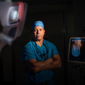
Flight-simulation technology used by military pilots to rehearse complex flight maneuvers is now helping neurosurgeons at NYU Langone to practice critical missions of their own: removing brain tumors and repairing aneurysms. The adapted technology, called the SuRgical Planner™, or SRP, combines MRI and CT scans to create detailed three-dimensional computer models of a patient’s brain. Using a joystick, surgeons manipulate the computer model on-screen so that they can see exactly where a tumor sits in relation to surrounding arteries, veins, and brain structures before they operate.
The developers, a neurosurgeon and a pair of Israeli military engineers, paid special attention to the simulator’s realism. Tissue glistens in the light and responds with lifelike movement to virtual surgical tools. “The SRP lets us walk around a tumor, in a sense, and see what’s in the way so that we can plan the safest approach for surgical resection,” explains John G. Golfinos, MD, chair of the Department of Neurosurgery.
Since its debut at NYU Langone this year, the FDA-approved surgical simulator has helped neurosurgeons rehearse for dozens of challenging cases, including ones in which tumors are nestled at the base of the skull, dangerously close to blood vessels, nerves, and brain structures. “Before, we had to do all the visualizing in our minds,” says Dr. Golfinos. “That’s not the same thing as a 3-D model, no matter how good you are. Neurosurgeons always spend extra time before every complicated case with the MRI and CT scans, looking for small but critical details. With the SRP, it’s almost as if you’re inside the MRI yourself.”

