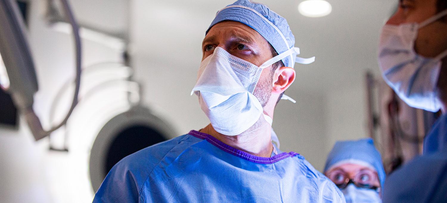
Neurosurgeon Dr. Donato R. Pacione, in collaboration with a multidisciplinary team of experts, deciphered a patient’s severe symptoms to identify—and safely remove—a rare pituitary tumor.
Photo: NYU Langone Staff
When a patient presented with a series of increasingly severe idiopathic symptoms, a multidisciplinary team of endocrinologists, neurosurgeons, and radiologists pursued an elusive diagnosis to identify her condition’s root cause: a rare pituitary tumor. Supported by advanced diagnostics and intraoperative imaging, the team collaborated to plan a minimally invasive surgical resection—and halt the patient’s decline.
Accurate Diagnosis Requires Extensive Diagnostic Sleuthing
The 38-year-old patient had a complex history, with a range of comorbidities including prior stroke, hypertension, cardiomyopathy, and treatment-resistant type 2 diabetes mellitus with hyperglycemia. Despite several hospitalizations at other centers, her symptoms continued to elude a definitive diagnosis. She consulted with a multidisciplinary team at NYU Langone, including endocrinologist Eliud Sifonte, MD, that suspected and confirmed hypercortisolism and a right-sided adrenal adenoma thought to be benign and unrelated to the hypercortisolism. Indications pointed to Cushing’s syndrome. However, the source of the hormonal irregularity remained unconfirmed, and the patient’s symptoms persisted, threatening her life and demanding swift diagnosis.
“With a case like this, it’s critical to really nail down the diagnosis and determine what is essentially giving this 38-year-old patient the clinical profile of a sick 75-year-old,” says Nidhi Agrawal, MD, clinical assistant professor in the Department of Medicine and director of pituitary diseases at the Pituitary Center. “And that diagnosis necessitates far more than a single blood test in isolation—you need a combination of clinical suspicion and the right advanced tools to confirm it.”
“With a case like this, it’s critical to really nail down the diagnosis and determine what is essentially giving this 38-year-old patient the clinical profile of a sick 75-year-old.”—Nidhi Agrawal, MD
In this case, clinical suspicion focused on the patient’s pituitary gland. Physicians used a combination of analytical tools including the highly sensitive adrenocorticotropic hormone (ACTH) assay, which is offered at few centers outside NYU Langone. This assay is critical to diagnostic specificity.
With diagnostic indicators pointing almost certainly toward pituitary involvement, Donato R. Pacione, MD, assistant professor in the Department of Neurosurgery, collaborated with Eytan Raz, MD, PhD, assistant professor in the Department of Radiology, to perform inferior petrosal sinus sampling, which would confirm the diagnosis. Response from the pituitary to microcatheter-injected hormone confirmed it was the source of the patient’s symptoms, and imaging subsequently revealed a pituitary microadenoma, a rare type of pituitary tumor.
Resecting the Root Cause, Carefully
Diagnosis in hand, the surgical planning began. “After the long road to diagnosis, surgery should be the simpler part—except that you’re working with something that’s 6 millimeters in size, and if you don’t resect every trace of the tumor, it still secretes hormone,” notes Dr. Pacione.
Understanding the historically high recurrence rate of such pituitary tumors, Dr. Pacione recommended an endoscopic endonasal approach for tumor resection, a minimally invasive approach that would enable precise navigation. Dr. Pacione worked closely with Elcin Zan, MD, assistant professor in the Department of Radiology, to develop a detailed anatomical map that would inform his surgical target.
During the procedure, the approach was performed by Seth M. Lieberman, MD, assistant professor in the Department of Otolaryngology—Head and Neck Surgery. The gland was identified and elevated to reveal inferiorly the tumor tissue, which Dr. Pacione dissected and removed. He also observed two other firm nodules anterior to the gland, in an area Dr. Zan had identified as a potential site of additional abnormal tissue, and removed them. When an intraoperative MRI demonstrated no evidence of residual tumor, the patient returned to the operating room (OR), where Dr. Lieberman performed the closure, and then moved to recovery.
Along with the careful multidisciplinary planning, advanced intraoperative imaging was critical to confirming full resection of the tumor. The intraoperative MRI, stereotactic navigation, and an advanced intraoperative histology system—which quickly and accurately identifies residual tumor while sparing healthy tissue—worked in tandem in a surgery that demanded extreme precision to achieve a positive outcome.
“Real-time, intraoperative feedback is the game changer—it enables us to both make faster surgical decisions and leave with a higher degree of confidence that the tumor is completely out,” observes Dr. Pacione.
Expertise and Access Change Clinical Trajectory
Within a day of surgery, the patient’s hormone level normalized, and she lost 30 pounds in 4 weeks. For this patient, multidisciplinary coordination of care, access to leading-edge diagnostic tools and imaging, and a collaborative pursuit of differential diagnosis reversed a precipitous decline in health toward a better outcome.
“This patient was already in heart failure and probably would have died if we hadn’t pinpointed and corrected her root issue,” notes Dr. Pacione. “It’s the difference between care that simply solves whatever is immediately broken on a given day and finding the underlying cause. That’s what changes the course and saves a patient.”

