Judy W. Lee, MD
PHOTO: Karsten Moran
When a complex presentation of facial paralysis and hearing loss led NYU Langone physicians to uncover a patient’s rare tumor, a multidisciplinary surgical team mobilized to coordinate its careful resection—and restore the patient’s function and appearance.
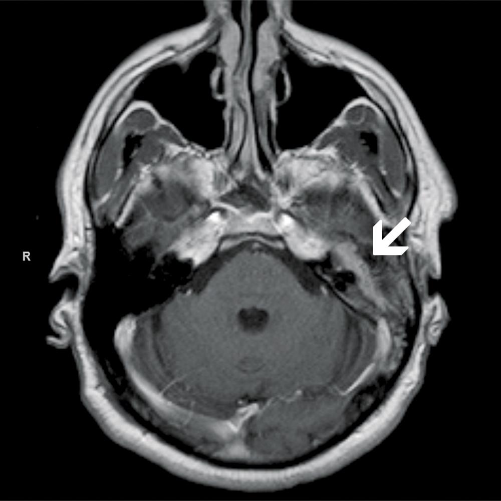
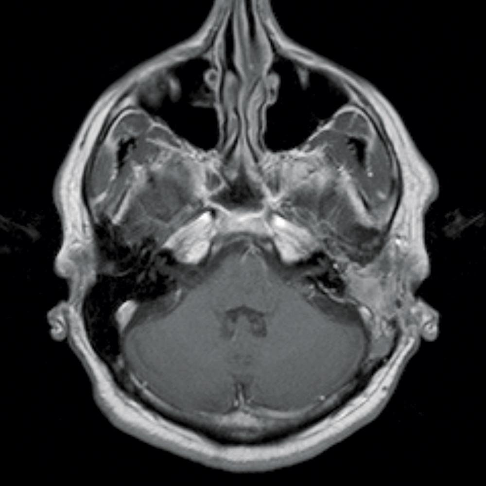
A 49-year-old man previously diagnosed with Bell’s palsy presented with persistent facial weakness and continued hearing loss with intermittent tinnitus. Judy W. Lee, MD, clinical assistant professor of otolaryngology—head and neck surgery, performed the initial exam, which showed complete facial nerve paralysis on the patient’s left side, with an ipsilateral middle ear lesion. Dr. Lee referred the patient for imaging tests and further evaluation, which led to a diagnosis of a large facial nerve schwannoma.
“Tumors of the facial nerve often cause facial paralysis, but they are fairly rare and often go undetected in the early stages,” says Dr. Lee. “Imaging showed a tumor extending along the patient’s facial nerve and filling his entire ear, causing significant hearing loss on his left side.”
Intracranial Tumor Excision Requires Skull Base Expertise
With the diagnosis confirmed, Dr. Lee referred the patient to Daniel Jethanamest, MD, clinical assistant professor of otolaryngology—head and neck surgery and director of the Division of Otology–Neurotology, and Donato R. Pacione, MD, clinical assistant professor of neurosurgery, who worked together to excise the tumor. The surgeons used a lateral approach in combination with a traditional microscopic approach with endoscope-assisted dissection to follow the full course of the lesion, excising the extensive schwannoma, which extended from the skull base to the stylomastoid foramen of the temporal bone. The patient had experienced nearly complete conductive hearing loss as a result of to the tumor’s erosion of the ossicular chain and its extension into the middle ear, which the surgeons also reconstructed during the surgery.
“Most of the pathology was in the temporal bone, but the tumor extended intracranially, and our biggest challenge was in reaching that cranial component of the tumor,” says Dr. Jethanamest. “This was a true team effort—an endoscopic-assisted approach allowed us to avoid the need for a larger craniotomy.”
The Tumor Excised, the Team Turns to Reanimation and Restoration
After the resection, the patient was evaluated for facial reanimation surgery by Adam S. Jacobson, MD, clinical associate professor of otolaryngology—head and neck surgery and associate director of the Division of Head and Neck Surgery, and Jamie P. Levine, MD, associate professor of plastic surgery and chief of microsurgery. Dr. Levine and Dr. Jacobson created a plan to achieve reanimation with a left gracilis free flap innervated by the left masseteric nerve, as well as a cross-face nerve graft from the buckle division of the contralateral side to the gracilis flap via a sural nerve graft, supercharged with two superior labial sensory nerves.
“During this surgery, we located the contralateral buccal branch of the facial nerve, which innervates many of the muscles of facial expression, including the levator anguli oris, levator labii superioris, and orbicularis oris,” says Dr. Jacobson. “We then connected the contralateral buccal branch of the facial nerve to the gracilis flap using the sural nerve graft, which was intended to give the patient more spontaneous movement and control over time.”
Dr. Levine performed a facelift to tighten the skin and a static sling of the left face to restore symmetry. Dr. Lee then implanted a 1.6-gram platinum weight so that the patient could close his left eye.
Improved Hearing—and Quality of Life
Since the procedure, the patient’s hearing has improved significantly across low to midrange frequencies and he has recovered excellent speech discrimination. His eye has regained full closure, and his facial swelling has been reduced. He does not yet exhibit facial movement, but on the basis of a very similar, successful reanimation case, physicians expect some movement will return within six months of the procedure, with continued improvement over time.
“Not only did our multidisciplinary team approach lead us to identify and remove this rare tumor that had eluded diagnosis for nearly a year,” says Dr. Lee, “but with a carefully coordinated, multispecialty surgical plan, we also recovered much of the function the tumor had impaired—and restored this patient’s quality of life.”


