

Main content
Our heart specialists use several tests to diagnose an arrhythmia before a baby is born, during infancy, or later in childhood.
An arrhythmia occurs when the heart’s electrical system, which sends signals that tell the heart to contract, isn’t working properly. This may cause the heart to beat too fast, which is known as tachycardia, or to beat too slowly, which is called bradycardia. Other types of arrhythmias can also make the heart beat irregularly.
Normally, a baby’s heart beats 100 to 150 times a minute. After a child’s first year, resting heart rate usually declines steadily until just before adolescence, when it levels off to about 55 to 100 beats a minute.
Arrhythmias that occur during infancy and childhood can be temporary or permanent. Babies and children with congenital heart disease or a family history of arrhythmias are at higher risk for an arrhythmia. So are those with certain genetic syndromes, such as familial dysautonomia, which affects the portion of the nervous system that directs involuntary functions, such as breathing and heart muscle contraction.
Conditions that cause inflammation in the heart, including infections of the heart and autoimmune conditions, can lead to an arrhythmia. Medications used to treat attention deficit hyperactivity disorder, chemotherapy drugs, and excessive amounts of caffeine can also cause one.
Symptoms of an arrhythmia in babies may include feeding problems, difficulty breathing, or excessive sweating. Parents should seek immediate medical attention if their baby has a sudden change in alertness or if the baby stops breathing and turns blue.
Symptoms in children may include palpitations, which make the heart feel as if it’s fluttering or pounding; dizziness, which can lead to fainting; and fatigue, especially during physical activity.
Heart specialists at Hassenfeld Children’s Hospital at NYU Langone use a variety of tools to identify an arrhythmia before and after a baby is born, as well as later in childhood.
An arrhythmia diagnosis may begin before a baby is born if the doctor notices an unusual heart rhythm during a routine prenatal ultrasound exam.
Doctors may also recommend prenatal monitoring of a baby’s heart rhythm if the mother has an autoimmune disorder, which could cause antibodies to block signals in the baby’s electrical system, or if a parent or sibling has congenital heart disease.
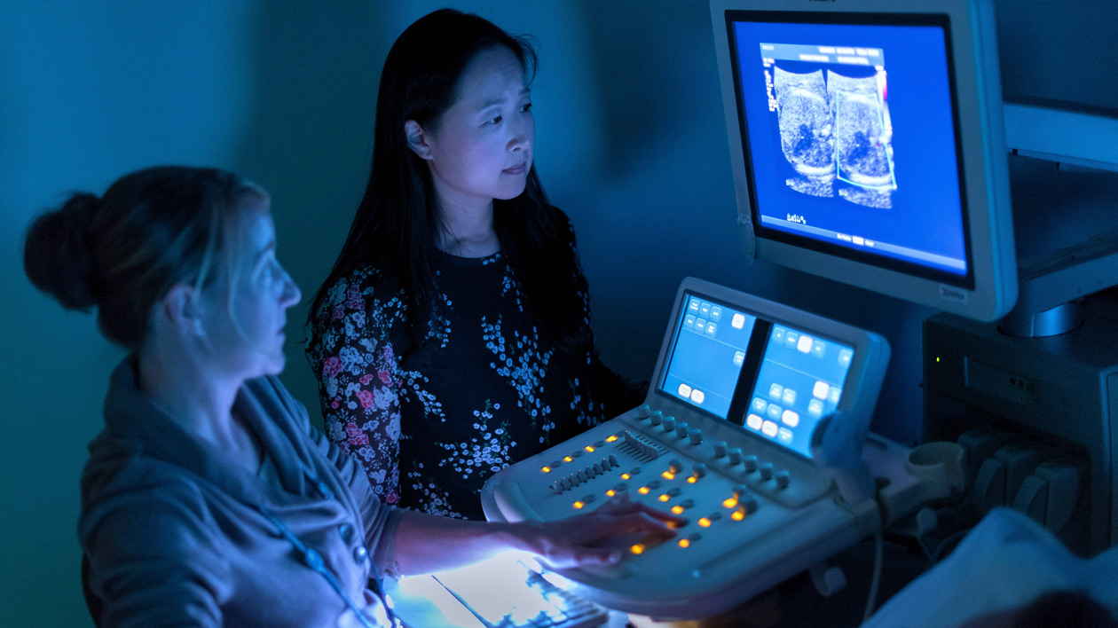
During a fetal echocardiogram, a doctor places a handheld ultrasound device called a transducer on the mother’s abdomen or inside the vagina. The probe transmits sound waves that bounce off of the baby’s heart. The sound waves are converted into a computerized image that reveals a picture of the baby’s heart and records heart rhythm.
If prenatal tests reveal that your baby may have an arrhythmia, your obstetrician can refer you to NYU Langone’s team of maternal–fetal medicine specialists for regular monitoring of your baby’s heart. If the arrhythmia becomes severe during pregnancy, our doctors can recommend treatments that can reach the baby through the placenta, the temporary organ that forms the baby’s life-support system during pregnancy.
At Hassenfeld Children’s Hospital, heart specialists use a physical exam and other tests to diagnose arrhythmias in babies and children or to confirm a suspected arrhythmia in babies with a known congenital heart defect or other risk factors. Many of these tests also help our doctors to determine the severity of the condition.
During a physical exam, the doctor uses a stethoscope to listen to your child’s heart. The doctor may also ask you or your child about any symptoms, such as feeling lightheaded or fainting.
An electrocardiogram, or EKG, is a test that records electrical activity in the heart. To perform the EKG, a technician places 10 stickers that contain small electrodes on your child’s chest, arms, and legs. The electrodes conduct electrical impulses from the heart to a machine that traces electrical activity, recording your child’s heart rhythm.
Your child must remain very still and quiet for a few minutes while the test is performed, but no sedation is needed, even in babies. Depending on what the tracing shows, the EKG can help the doctor determine whether the pattern of electrical activity is normal.
The doctor may also ask your child to exercise on a treadmill or stationary bicycle to determine whether physical activity causes the heart rhythm to be irregular.
A Holter monitor is a portable device that can be used at home to record your child’s heart rhythm over a 24-hour period, during everyday activities, and while your child is sleeping. The device contains a small, rectangular recorder that can be worn around the waist. The recorder is connected to small electrodes that are stuck to your child’s chest.
An event monitor is another portable device used to measure electrical activity at home for a week or longer. Unlike the Holter monitor, which records electrical activity continuously, the event monitor only records electrical activity when your child experiences symptoms.
A technician can explain how and when to push a button that instructs the monitor to record electrical activity and how to send the data to the doctor’s office.
An implantable monitor may be used to record heart rhythm for up to three years in children who have a high risk of developing an arrhythmia or have sporadic symptoms, such as fainting, that could be caused by an arrhythmia. Doctors place this small device, about the size of a flattened pen cap, just under the skin of the chest.
Surgery to implant a heart monitor is performed in the hospital and requires anesthesia. General anesthesia is usually preferred for young children, but local anesthesia may be used for adolescents. The procedure takes about 30 minutes, and most children can return home the same day.
Our doctors determine how frequently to check the heart monitor based on the severity of your child’s symptoms. Data from the monitor can be retrieved in the doctor’s office or at home, using a wand that is held on the chest, above the monitor, to transmit data to the doctor’s office.
A transesophageal echocardiogram is an imaging test that uses ultrasound to create an image of the heart. In this test, the doctor guides a small probe through the nose and into the esophagus—the tube through which food travels from the throat into the stomach—to the upper part of the chest near the heart.
This test, which is performed in less than an hour using mild sedation, can provide a more detailed image of the heart than a conventional echocardiogram performed on the surface of the chest.
During an electrophysiology study, which requires anesthesia, a doctor inserts small, thin wire electrodes through a vein in the groin or neck and threads them into the heart. The heart rhythm physician uses a special magnetic device to map an image of the heart and to precisely identify the location of the wires with little or no radiation.
Electrical signals are sent to the heart via the electrodes to see how the heart responds. This helps to determine whether the arrhythmia is caused by a problem with the heart’s electrical system.
An electrophysiology study is more invasive than other tests for arrhythmia. For this reason, it is usually done only when the doctor needs to determine whether the child is at risk for a life-threatening event.
The doctor may also perform this procedure if he or she suspects the child has an electrical problem that can be treated during this test. If such a defect is present, the doctor can perform a minimally invasive procedure called catheter ablation to correct the cause of the arrhythmia.
Information from these tests can help our doctors to determine which treatment your child may need and when treatment should begin.