

Main content
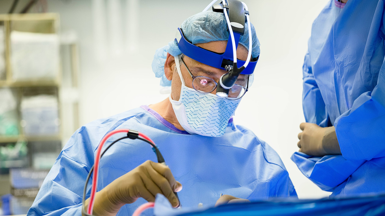
Surgeons at Hassenfeld Children’s Hospital at NYU Langone use surgery to correct congenital abnormalities that affect a child’s airway. This includes procedures to widen narrowed airways, reduce swelling, create openings to the larynx, or voice box, and remove obstructions, such as irregular tissue or lesions. Some procedures can be performed at the same time as diagnostic tests, such as during a triple endoscopy.
Our surgeons use techniques such as laser surgery to remove abnormal tissue or cysts. They may also use minimally invasive endoscopic surgery to reconstruct abnormal organs or cartilage and to perform mucosal grafting, in which a piece of cartilage is removed and surgically attached to keep the trachea open.
Sometimes surgery is performed while a baby is in the womb. In these situations, the mother is given medications to prevent uterine contractions during surgery, which can result in premature delivery. This surgery is performed with general anesthesia.
Experts at the Pediatric Aerodigestive Center, part of Hassenfeld Children's Hospital, also provide tracheostomy care for children who have difficulty breathing. This includes the insertion of a tube into the trachea to allow your child to breathe adequately. Doctors also determine if your child is ready for decannulation, in which the tracheostomy tube is removed so your child can breathe naturally.
In a tracheostomy, a surgeon inserts a plastic tube directly into the trachea, or windpipe. In some children, this tracheostomy tube is connected to a ventilator machine, which supports breathing by supplying oxygen to the lungs and removing carbon dioxide from the body.
During this procedure, which requires general anesthesia, the surgeon makes an incision in the neck and inserts a plastic tracheostomy tube, which is held in place with surgical tape or sutures.
If needed, our specialists can use a three-dimensional printer to custom fit a tracheostomy tube to the unique anatomy of your child’s airway. This allows doctors to better fit the tube into the airway.
After this procedure, your child remains in the hospital for a week. Our experts teach you how to care for the tracheostomy tube before you go home.
Children who no longer need a tracheostomy tube to assist in breathing may have it removed in a procedure called decannulation.
Our specialists first check the child’s airway for obstructions using laryngoscopy or bronchoscopy. Next, a cap is placed over the tracheostomy tube to assess the child’s ability to breathe through the mouth and nose. The cap is removed if there are signs of breathing difficulties.
The tube is then capped overnight while the child sleeps. His or her heart rate, breathing, and oxygen levels are monitored. If there are no difficulties, the tracheostomy tube is removed the next day, and the incision is covered. Your child remains in the hospital for 24 to 48 hours for observation.
Your child returns to the doctor four to six weeks after decannulation, so that he or she can check that the incision is healing well. The doctor also ensures that your child isn’t having any problems with his or her voice, such as hoarseness.
Anti-reflux surgery may be performed to treat children who have severe and complicated gastroesophageal reflux disease, which is when acid, liquids, and food in the stomach rise into the esophagus and throat and often drip into the trachea and lungs. This can cause a range of symptoms from sore throat and heartburn to aspiration pneumonia, which is an infection of the lungs.
During this procedure, which requires general anesthesia, the surgeon wraps part of the stomach around the bottom end of the esophagus to help tighten muscles and prevent stomach acid and food from rising from the stomach. Or the surgeon may widen the opening between the stomach and the small intestine, allowing the stomach’s contents to empty faster.
Our surgeons can perform these procedures by making an incision in the abdomen—called open surgery—or by using a laparoscopic technique, in which several small incisions are made in the abdomen. Surgical tools and a laparoscope, which is a thin tube with a tiny camera at its end, are inserted into these incisions. The surgeon then uses these tools and the assistance of a computer to perform the procedure.
Your child may remain in the hospital for two to three days after this procedure, or longer if he or she had open surgery. Sometimes a gastrostomy tube, or G-tube, is placed in the abdomen to provide liquid nutrition to the child.